Columbia Engineers Develop Implantable Imaging Tool to Reveal Deep-Brain Neural Circuits
A team led by Ken Shepard unveils a high-precision platform that opens new possibilities for probing the brain's deep circuits.
05 Mar, 2026.
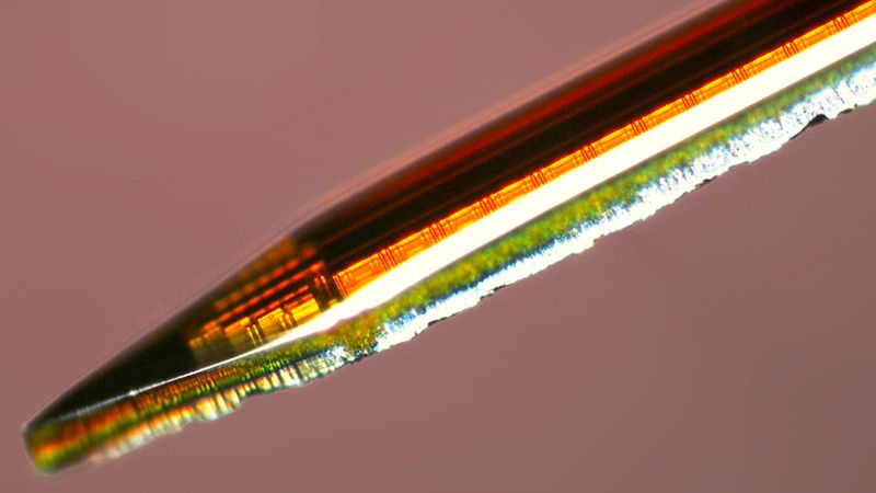
Acus is a CMOS implantable fluorescence imager to monitor neural circuits in deep regions of the brain. Credit: Bioelectronics Systems Lab at Columbia University
This article was first published on
www.engineering.columbia.eduA Columbia Engineering team has introduced a powerful new tool for neuroscience, offering researchers a new way to monitor neural circuits in deep regions of the brain. The work is led by Kenneth Shepard, Lau Family Professor of Electrical Engineering, professor of biomedical engineering, and professor of neurological sciences (in Neurosurgery) at Columbia University. The lead authors of the study, published in the December issue of Nature Electronics, are PhD students Sinan Yilmaz and alumnus Jaebin Choi PhD‘21.
The paper presents Acus, an ultra-miniaturized neuroimaging platform capable of reaching brain regions that traditional optical tools struggle to access. The system consists of a microscale complementary metal-oxide-semiconductor (CMOS) probe that integrates an array of single-photon avalanche diodes (SPADs), spectral filters, and an optical fiber for light delivery to create a compact implantable fluorescence microscope in a needle-like form factor. While similar CMOS platforms are used for electrophysiological interfacing, Acus moves these same techniques into the optical realm. This allows cell-type-targeted imaging at depth while maintaining a footprint small enough for chronic implantation with minimal tissue disruption.
In conventional fluorescence imaging, light scattering and absorption in brain tissue severely restrict imaging depth, confining observations largely to superficial cortical layers and limiting access to deeper neural circuits. By implanting the microscope itself, Acus enables precise volumetric localization of individual neurons at depths beyond those achievable with conventional microscopes, allowing researchers to reliably monitor neuronal activity deep in the brain while preserving the animal’s natural behavior.
“Optical imaging modalities have significant advantages over electrophysiology including precise localization and cell-type specificity,” says Yilmaz. “We think that Acus is a valuable new tool for neuroscientists to study neural circuits with single-cell resolution. We have already started several new collaborations with neuroscience labs across the country.”
The work underscores Columbia Engineering’s leadership at the intersection of electronics, biology, and neurotechnology. Shepard’s lab, the Bioelectronic Systems Lab, has long advanced high-density neural interfaces and implantable electronics leveraging the scalability of CMOS process, and this latest publication introduces another new neurotechnological tool leveraging leading-edge microelectronics and optical packaging.
The work was done in collaboration with Michael Roukes at the California Institute of Technology.